
Preparing practitioners for the future
Beginning with an early push into digital x-rays, as well as having the first comprehensive electronic patient record chairside in a dental school, the UMKC School of Dentistry has long instilled an innovative spirit in their students. Today, that continues with the school’s drive into the era of modern dentistry.
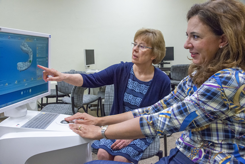
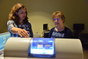
The latest addition to the school’s digital dentistry offerings are 3D printers – devices used to “print” (or make) everything from clear orthodontic aligners and dentures to surgical guides for implant cases. These new printers are part of the school’s current digital equipment lineup, which includes state of the art treatment planning software, intra and extra oral scanners, cone beam tomography (CBCT) and milling systems for restorations. And for several years, the school has been providing students experience in creating and using digital impressions in laboratory simulations, as well as patient care.
“Having these tools in place and getting students comfortable and familiar with using them will give them a huge advantage moving forward.”
Dr. Cynthia Petrie, Chair of Restorative Clinical Sciences
It’s this extremely rapid evolution and confluence of technology, lack of standards, potential for duplication of resources and high costs of care that led Dean Marsha Pyle to establish the school’s Digital Dentistry Task Force. “This technology covers a wide spectrum of departments at the school,” said Pyle. “Our new task force brings all the players to the table in one forum to develop a road map for future planning.”
Scanning to 3D printing- digging into digital
For Dr. Mary Walker, Associate Dean for Research and Graduate Programs, the acquisition of the 3D printer, in particular, is a great example of the need for and benefit of collaboration. According to Walker, many times in private practice 3D printers are used for very specific purposes, such as orthodontics. But at UMKC’s dental school, the printer would need to cross many different disciplines and uses.
“When we purchase this type of equipment, it’s critical that we look at multi-users and multiple applications,” said Walker. “We tried to bring together the departments and disciplines to meet everyone’s needs.”
Of the 3D printers at the school, one is a high-end, precision printer to be used for very precise and complex procedures, such as “printing” (producing) scaffolds for bone tissue regeneration to treat craniofacial defects that can occur genetically, or from trauma or disease. For example, the school has researchers working with surgeons to try and build orthopedic components to augment or replace bone where a patient has lost part of their maxilla. Using the printer to create a scaffold, doctors would be able to a replicate and implant the patient’s jaw structure on which to grow new bone.
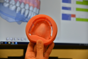
The school also has two other printers that produce more traditional dental applications, such as clear orthodontic aligners, dentures and crowns.
Walker, who has been teaching and researching using Digital Dentistry at the school since 2002, has seen many changes in the field over that time. But she says one aspect in particular has remained: enhancing the patient experience.
“I think at the end of day, the patient appreciates that their dentist – at the school or in private practice – is on the cutting edge of technology,” said Walker. “They want to be assured that we know what’s new and know how to use it.”
 According to Dr. Cynthia Petrie, Chair of Restorative Clinical Sciences, the school’s use of digital technology had humble beginnings. “In 2013, when we started teaching students how to mill crowns and design them digitally, we were using a space that was shared with yoga classes. But we were persistent on the importance of teaching digital dentistry to the students. Now, we have a robust curriculum in which each student receives instruction in digital dentistry in all four years of the dental curriculum. They then use this technology when providing prosthodontic treatments for their patients.” Petrie said.
Although the school introduced digital scanning for dental impressions a couple years ago, currently both conventional and digital techniques are taught for prosthodontic treatments in the patient care clinic. The students get experience making conventional 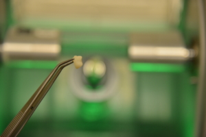 impressions too, seeing the advantages and limitations of both methods. Beyond digital impressions, students learn to design and mill crowns and bridges from the digital impression scan files, plus they receive experience with the digital design and fabrication of dentures.
Since the school has been providing this training for a couple of years, Petrie is studying the long-term data on the success rates of digital dentistry in the predoctoral clinical curriculum. To date, when comparing outcomes to other schools and published reports in the literature, the data from UMKC School of Dentistry has been quite favorable. Petrie attributes this success to the extensive experience the students receive in pre-clinic courses, mastering basic skills and processes via simulation and rigorous instructions from faculty and teaching assistants.
“We think that the didactic and preclinical curriculum prepares the students adequately to be able to use these techniques and technology effectively and efficiently for patient care in the D3 and D4 years,” said Petrie.
Putting digital into practice
Dr. Pete Spalitto (D.D.S. ’00) graduated ahead of the school’s push into digital impressions. But he says his experience with the school’s clinical innovative stuck with him, and still holds true in his practice, as well. “When it comes to what technology we’ve implemented in my practice,” said Spalitto, “it’s better to ask what we haven’t put in place.”
“When my staff is empowered to take ownership of the technology, it just makes us that more powerful of team.”
Dr. Pete Spalitto (D.D.S. ’00)
Through the years, his practice has added a diode laser, two milling machines (chambers), digital X-rays and CBCT scanner, among other things. Spalitto says the CT scanner has been an incredibly helpful addition for his patients. “I take a CT scan of every root canal I’m doing now,” he said. “It’s like giving me a GPS map to the framing of the nerve endings.”
It’s also been important for Spalitto to bring the rest of his staff in early when integrating any piece of new technology. “When my staff is empowered to take ownership of the technology, it just makes us that more powerful of team,” he said.
According to Spalitto, his time at the school laid the foundation for his technology adoption. “I think the role of any health professions school is to give you the tools to succeed, but also to get you thinking outside of the box,” he said. “My professors were always discussing different technology and encouraging us to integrate it into our future practices.”
Learning together through technology
Everyone agrees it’s important that the students and faculty learn this technology together. Even though today’s students have grown up with technology all around them, there is still a learning curve to grasping the industry’s unique digital experience, selecting the most appropriate solution and using it most effectively for the patient’s care.
“This is where dentistry is headed in the future and it’s going at a very fast pace,” said Petrie. “Having these tools in place and getting students comfortable and familiar with using them will give them a huge advantage moving forward.”
