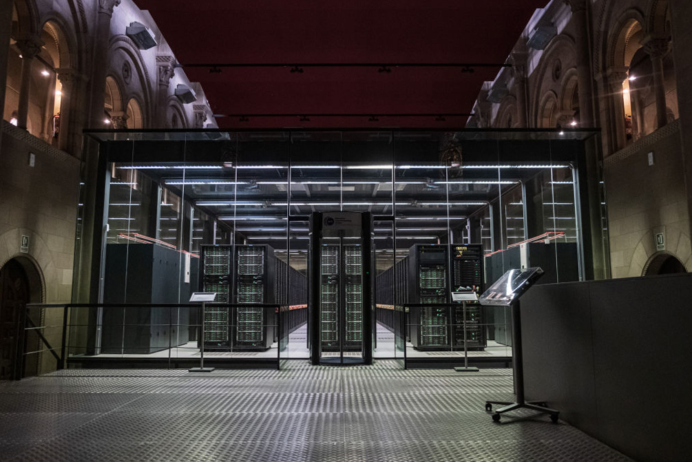
Within the walls of a 19th-century chapel on the outskirts of Barcelona, a heart starts to slowly contract. This is not a real heart but a virtual copy of one that still pounds inside a patient’s chest. With its 100 million patches of simulated cells, the digital twin—a fully functional simulation of human anatomy— pumps at a leisurely pace as it tests treatments, from drugs to implants.
This digital twin pulses within MareNostrum, a supercomputer used by scientists to simulate features of the real world. These simulations can look just like the real thing, but they are vastly more sophisticated than Hollywood visual effects because they behave like the real thing—from how the heart moves to the charged atoms that zip in and out of its cells. And they are already beginning to help doctors to predict how the particular heart of a particular patient will respond to a particular treatment.
This worldwide effort to create virtual cells, tissues, and organs is revolutionary because modern medicine is largely backwards facing: Currently, doctors try to figure out what is the best treatment for their patients by assessing past clinical trials on subjects who are a bit like their present patients, in similar but not identical circumstances. In coming decades, though, doctors will be able to use digital twins—which see the exchange of data and insights between a real and virtual human—to better predict what lies in store for patients, helping what is mostly a one-size-fits-all approach evolve into one that is truly predictive and personalized.
Read More: How Digital Twins Are Transforming Manufacturing, Medicine and More
Virtual organs are the culmination of research that stretches back more than half a century, to experiments in the 1950s on the conveniently-large nerves from a squid. That work helped Britons Alan Hodgkin and Andrew Huxley hone a groundbreaking mathematical theory on how nerve impulses propagate, which would later earn them a Nobel prize.
By 1959, a young English scientist Denis Noble began to apply Hodgkin and Huxley’s insights to heart cells. He realized it would take him months to use a hand-cranked calculator to work out what happens over half a second in a single cardiac cell, and, as a result, turned to the Ferranti Mercury, a valve computer in Bloomsbury, London, that weighed one ton and could do 10,000 floating point operations every second (the MareNostrum manages one thousand million million). After a few weeks, Noble plotted out the results from a teleprinter and was amazed to see that his virtual heart cell was “beating.”
Noble, who moved to Oxford in 1963, would develop his heart cell model with colleagues, such as New Zealander Peter Hunter. By the 1980s, they had a reasonable understanding of the electrical, chemical, and mechanical activities involved in the contraction of a single cardiac cell, boiled down to around 30 equations. At the end of the decade, working with American biomedical engineer Rai Winslow , Noble started work on a whole organ model of the heart on a Connection Machine, one of the world’s first commercial supercomputers based on parallel processing. This paved the way for digital heart twins.
Today, in Oxford, Computational Medicine Professor Blanca Rodriguez’s team has passed another important milestone for digital heart twins. While Noble’s first cell model relied on a handful of equations, her model uses several dozens. Most important, her human virtual heart predictions are more accurate than comparable animal studies, offering a way to reduce vivisection—the process of operating on live animals for scientific research. In one virtual “drug trial,” for example, where 62 drugs and reference compounds were tested in more than a thousand simulations of human heart cells, her team predicted the risk that drugs would cause abnormal heart rhythms with 89% accuracy. When they compared these computer predictions with data obtained from previously-conducted comparable animal studies, the animal research was less accurate (75%).
Similarly, scientists at the Barcelona Supercomputer Center have developed their Alya Red digital twin heart model, which consists of around 100 million patches of virtual heart cells, each of which is described by around 50 equations. For its most detailed, high resolution cardiac digital twins, Alya Red can take 10 hours to simulate 10 heart beats. The “blood” coursing through it can be brought to life as bundles of vivid colors with red, orange, and yellow revealing vigorous flows; the flows within diseased ventricles are revealed as sluggish blues and greens. In this way, the digital twin can reveal how a failing heart loses its ability to pump, or the dangerous arrhythmia caused by heart drugs. Working with the medical technology company Medtronic, the Alya Red simulations can help position a pacemaker, fine tune its electrical stimulus, and model its effects.
Examples of digital heart twins can be seen around the world. Led by Reza Razavi at King’s College London, for instance, digital twin heart models based on patient data have been created to predict tachycardia, an abnormally rapid heart rhythm that is a leading cause of sudden cardiac death. Meanwhile, at Johns Hopkins University, Natalia Trayanova’s team is creating digital replicas of the heart’s upper chambers to guide the treatment of irregular heartbeats by the targeted destruction of tissue and stop errant electrical impulses.
In France, the multinational software developer Dassault Systèmes (in partnership with the U.S. Food and Drug Administration) has created a cohort of “virtual patients” to help test a synthetic artificial heart valve for regulators. The FDA also used computer-simulated imaging of 2,986 virtual patients to study mammography that relies on low-dose x-rays—a huge feat because, until recently, regulatory agencies have relied on evidence from trials on people, rather than digital twins.
As a result, many scientists have begun to think about how to connect a virtual heart to a virtual body. In preparation for linking up to a virtual heart, our consortium, CompBioMed, has created a digital twin of a 60,000-mile-long network of vessels, arteries, veins, and capillaries that take blood around the body using billions of data points from digitized, high-resolution cross-sections of the frozen cadaver of a 26-year-old Korean woman, Yoon-sun, gathered as part of the Visible Korean Human project. We gained access to a German supercomputer, SuperMUC-NG for several days, which enabled us to model how virtual blood flowed for around 100 seconds through a virtual copy of Yoon-sun’s blood vessels down to a fraction of a millimeter across. Our team is now charting variations in blood pressure throughout the digital twin of Yoon-sun’s circulation and simulating the movement of clots.
In the U.S., a related effort is under way by computer scientist Amanda Randles at Duke University to study hypertension and how to treat cerebral aneurysmsbulges in artery walls—while, in Graz, Austria, Computational Cardiology Professor Gernot Plank is working with colleagues in the Netherlands and France to create a 3D model of the heart and circulatory system, which he told us is as close to a universal simulator of the heart’s electrical and mechanical behavior as anybody got so far.
Remarkable progress has also been made in creating digital twins of simple cells at Stanford University, while at the Auckland Bioengineering Institute, computer modeling pioneer Peter Hunter and his colleagues are working on an array of organs, from heart and lungs to liver and guts. Even the most complex known object, the human brain, is being simulated, for instance to plan epilepsy surgery in a French clinical trial. At last, the virtual human is heaving into view; in theory, it will help you better understand what forms of diet and exercise offer you a longer, happier life.
Along the way, we must be ready to deal with new ethical issues, of course: Our notion of what it means to be “healthy” may have to change. A person could be seen as “unhealthy,” suffering from a “symptomless illness,” or perhaps seen as being “asymptomatic ill,” if their digital twin shows that they could live significantly longer by changing their lifestyle, adopting a new diet, or using an implant or drug.
Ultimately, the rise of digital twins could pave the way for methods to enhance your body and your future. Virtual humans will hold up a mirror to reflect the very best that you can be.
More Must-Reads From TIME
- The 100 Most Influential People of 2024
- Coco Gauff Is Playing for Herself Now
- Scenes From Pro-Palestinian Encampments Across U.S. Universities
- 6 Compliments That Land Every Time
- If You're Dating Right Now , You're Brave: Column
- The AI That Could Heal a Divided Internet
- Fallout Is a Brilliant Model for the Future of Video Game Adaptations
- Want Weekly Recs on What to Watch, Read, and More? Sign Up for Worth Your Time
Contact us at letters@time.com